BESA Research 7.1
The BRAIN QUICK LTM Line offers easy expansion from 32 to 256 channels and can be used as a wireless, wired or ambulatory system. Its compact, lightweight footprint and limitless patient mobility makes the system ideal for pediatric LTM studies. The small, versatile SD LTM 32 PLUS and SD LTM 64 PLUS amplifiers can serve as a 3-in-1 system: wired headbox, wireless headbox or ambulatory recorder.
Choose the best signal processing for your EEG and MEG data
BESA is the most widely used software for source analysis and dipole localization in EEG and MEG research. BESA Research has been developed on the basis of 30 years experience in human brain research by the team around Michael Scherg, University of Heidelberg, and Patrick Berg, University of Konstanz.
BESA Research is a highly versatile and user-friendly Windows® program with optimized tools and scripts to preprocess raw or averaged data for source analysis and connectivity analysis. All important aspects of source analysis and source imaging are displayed in one window for immediate selection of a wide range of tools. The same holds true for the source coherence / time-frequency module, and other analysis windows. BESA Research provides fast and easy hypothesis testing, a variety of source analysis algorithms including cortical imaging and volume imaging methods, integration with MRI and fMRI, age-appropriate template head models (FEM) as well as the possibility to import individual head models (BEM and / or FEM) generated by BESA MRI.
Connectivity analysis, a unique feature for viewing brain activity
BESA Research transforms surface signals into brain activity using source montages derived from multiple source models or beamformer imaging. This allows displaying ongoing EEG/MEG, single epochs, and averages with much higher spatial resolution.
The BESA Connectivity solution and the Source Coherence Module provide an extremely fast and user-friendly implementation of time-frequency analysis based on wavelets and / or complex demodulation. Users can create event-related time-frequency displays of power, amplitude, or event-related (de)synchronization and several connectivity measures for the current montage using brain sources or surface channels. Induced and evoked activities can be separated. Source connectivity analysis reveals the functional connectivity between brain regions by reducing the volume conduction effects seen in surface coherence. Users can select a time-frequency window for (bilateral) beamformer and dynamic imaging of coherent sources (Gross et al., PNAS 2001).
More than just dipole source localization
BESA Research covers the whole range of signal processing and analysis from the acquired raw data to dynamic source images:
Features
Data review and processing module
BESA Research provides many tools for reviewing and processing of your EEG or MEG data. Raw data are directly read using readers implemented for many EEG and MEG file formats. Data processing steps include digital filtering, artifact detection and correction, computation of correlation and spectral analysis. All these steps can be performed easily with a few mouse clicks. A variety of different display options allows for convenient review of your data.
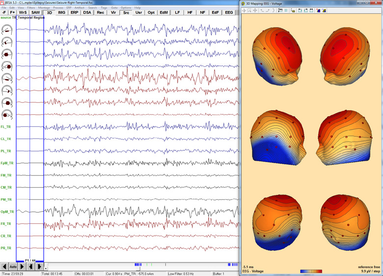
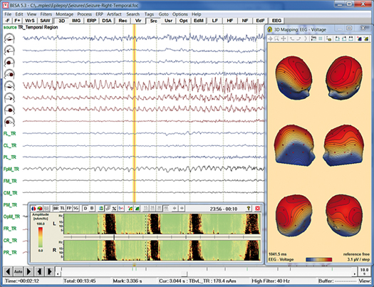
Onset of epileptic seizure with 3D whole-head maps
Data import and export
- Direct readers for most EEG and MEG data file formats
- Import of user-defined file formats using generic reader
- Data import / export to ASCII and binary files
- MATLAB interface for direct transfer of analysis results to MATLAB
Data processing
- Superior digital filtering: high, low, and narrow band pass, notch
- Interpolation from recorded to virtual and source channels
- Automated EOG and EKG artifact detection and correction
- Advanced user-defined instantaneous artifact correction
- Pattern detection and averaging by spatio-temporal correlation
Data review
- Easy and fast review of digital EEG and MEG data files
- Fast paging, tagging and selected viewing of epochs of interest
- DSA and event displays for quick jump to relevant pages
- Additional selected and virtual artifact channels (EOG etc.)
- Linear and non-linear correlation between scalp and source channels
- Spectral analysis: FFT, DSA, power and phase mapping
- Independent Component Analysis (ICA): Decomposition of EEG/MEG data into ICA components that can be used for artifact correction and as spatial sources in the source analysis window