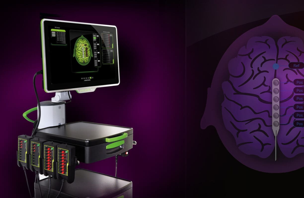
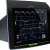
Neuromonitor for Spine & Neuro Surgery
AVALANCHE® PLUS
With the anatomical design of the user interface of our AVALANCHE® PLUS inspires its users and revolutionizes intraoperative neuromonitoring (IONM).
A consistent visual configuration guides the user and facilitates proper handling. The AVALANCHE® PLUS is intuitive to learn and easy to operate. This increases efficiency, saves valuable time.
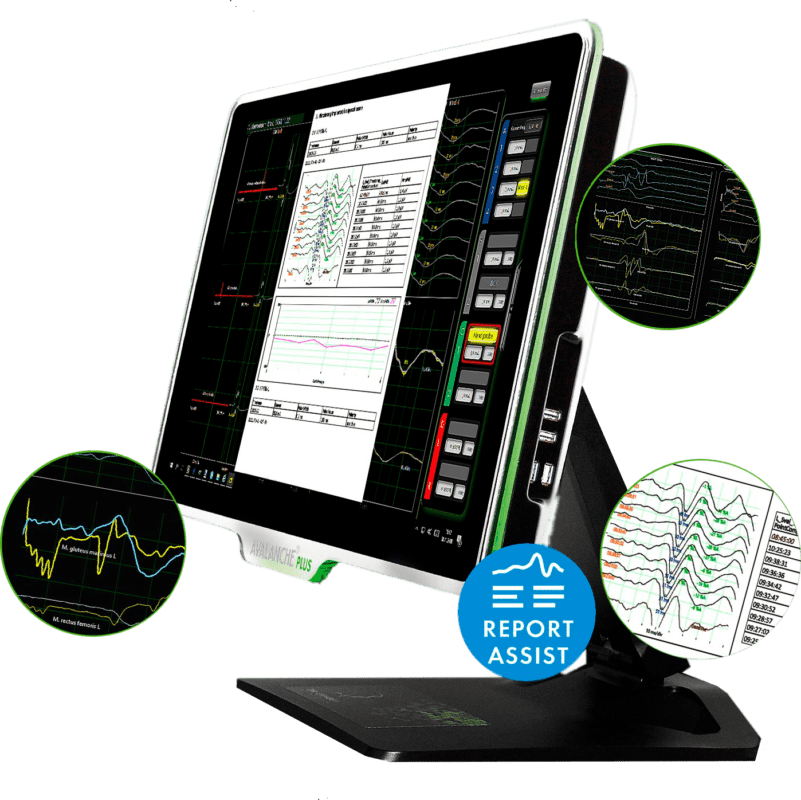
Perform the visual configuration completely via touchscreen, select individual muscle groups and assign them to the desired measurement window via DRAG & DROP, have a reliable view of the signals during the operation and then conveniently generate a PDF report. Exactly this kind of operating concept has never been implemented before in this field of medical technology.
The art of neuromonitoring – this is not only our slogan. The user interface graphics are objects of art that reflect our philosophy from the very beginning. This newly created user experience leads to more confidence in using the neuromonitoring device. We intend to bring more quality to the operating room with our devices with the goal of providing patients with even better care.
32-CHANNEL NEUROMONITOR
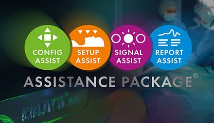
with ASSISTANCE PACKAGE®
Our new AVALANCHE® PLUS takes a completely new approach to operation. We call it ASSISTANCE PACKAGE®, because you are guided through the typical workflow of a surgery in four assisted steps. You will feel the advantages of our well thought-out concept. Just like a modern smartphone, all tasks are performed via touch screen. This is not only fun, but also promotes understanding of the complex multimodal neuromonitoring.
After a brief period of familiarisation, you will learn to appreciate the advantages of the system, you will need less time, errors will be found more quickly and largely avoided thanks to the ASSISTANCE PACKAGE® – our goal, more patient safety.

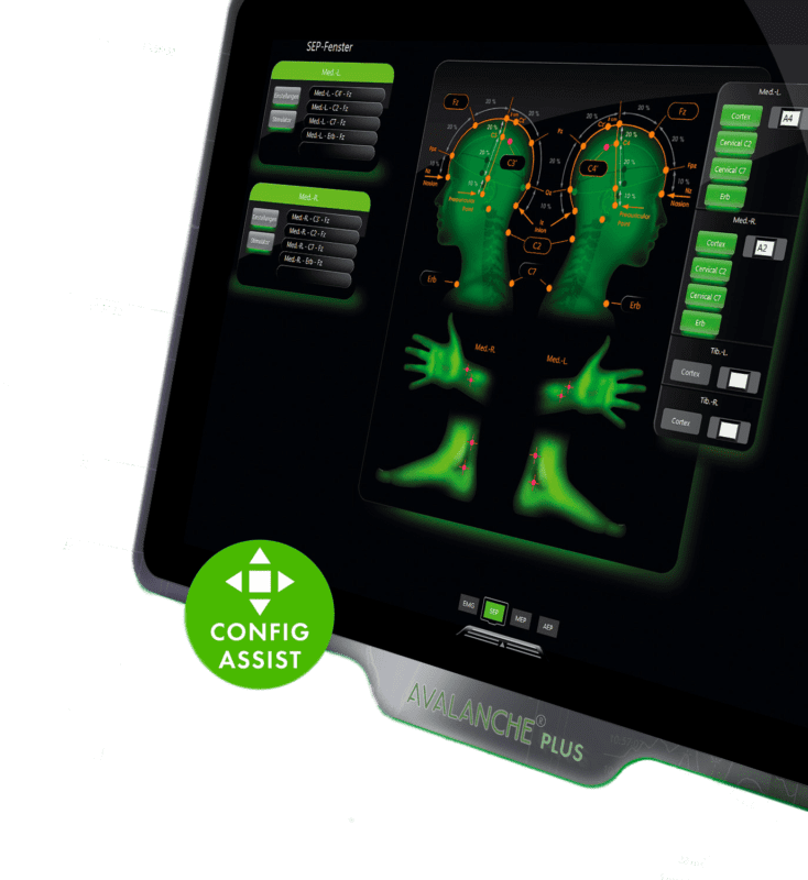
CONFIG ASSIST
You decide what you need
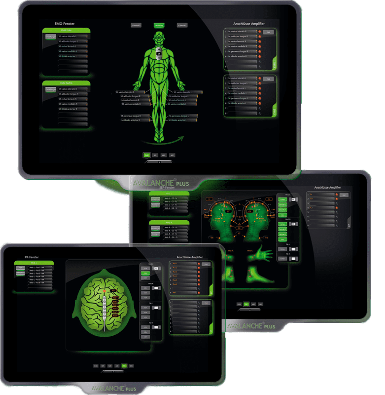
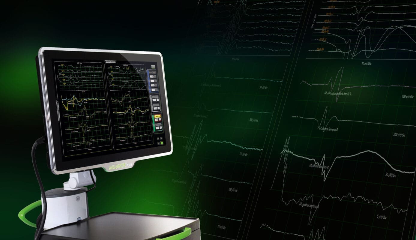
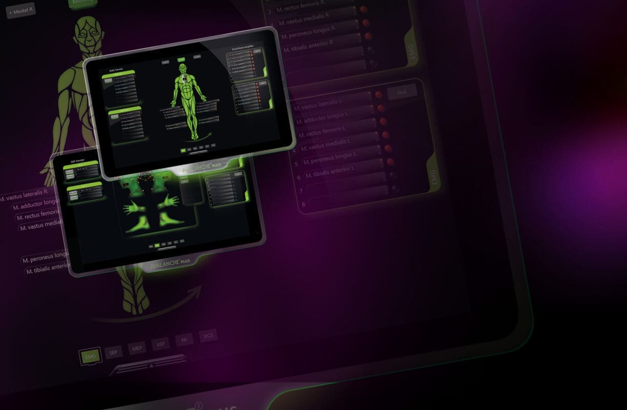
Handling our AVALANCHE® PLUS is guaranteed to be a new experience. You will master the system in no time at all. From the visual display, you determine your signal recordings in a flash. You control everything simply and clearly on the touch screen. No more thousands of functions and hidden screen windows that no one can remember.
Select individual muscle groups and assign them to the desired measurement window via DRAG & DROP. AVALANCHE® PLUS knows predefined recordings, e.g. for evoked potentials. Your measurement window is created at the touch of a button. The system automatically assigns the stimulators. You select connections for the electrodes and are ready to go. And the best thing: the system remembers all settings for you. With the help of markers, you can see at any time which recordings have been set – this is how quick it can be.
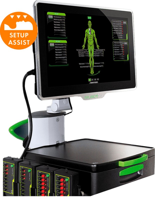
SETUP ASSIST
Patient connection made easy
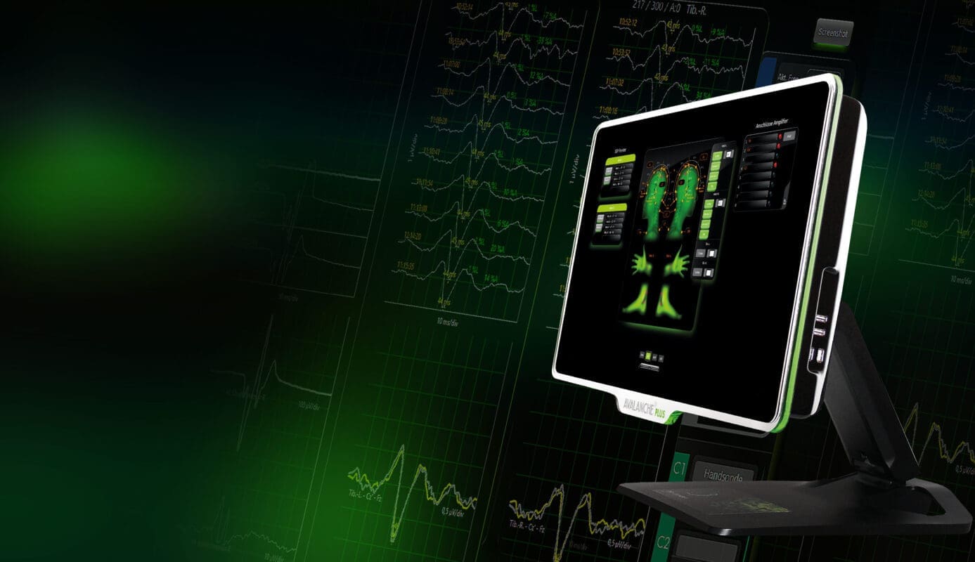
AVALANCHE® PLUS shows you at any time which electrodes are connected where or which connections are still missing. Our colour- and shape-coded connections, e.g. on our numerous stimulator outputs, can be quickly and clearly assigned.
AVALANCHE® PLUS also supports you in connecting the recording electrodes. When you select a muscle on the screen or a cortical recording position in the 10-20 system, a white LED on the amplifier near the patient shows you the correct connection. Not only can you find the exact electrode position immediately, but you can, for example, identify a bad electrode contact causing signal interference without significant loss of time. Because as soon as the electrode is placed,
AVALANCHE® PLUS continuously checks the electrode contact and even displays it directly on the preamplifier. Green for very good contact, yellow for sufficient contact and red for loss of contact – it could hardly be simpler.
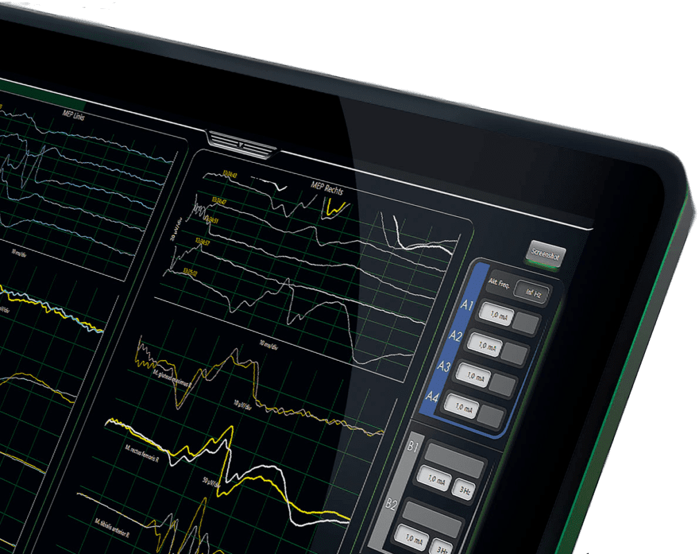
SIGNAL ASSIST
Never miss a thing
On the large touchscreen of the AVALANCHE® PLUS, you have all configured signals in view at all times. Separate views are also available for each modality – if the other modalities are not so important at the moment. RF interferences from electrosurgical units are automatically detected and faded out – without additional sensors that must be awkwardly attached. Recordings with unusuall high impedance are displayed in red, a click on the channel and the LED on the preamplifier shows the respective electrode. The error is quickly corrected and the signal can be recognised as “clean” again.
Stored measurement setups can be called up and used at any time. Via the quick access to the stimulators, you change the stimulation intensity for each stimulator directly from the main window and see its influence on the signal quality. Keep a full signal overview during the entire surgery to make sure you don‘t miss a thing.
MAKES YOU A SPECIALIST
See – Understand – Apply
Experience more of the ease with which you will perform neuromonitoring in the future. With our ASSISITANCE PACKAGE® in the new AVALANCHE® PLUS.
You will see the difference – we promise!
REPORT ASSIST
Final report at the touch of a button
Create screenshots in a single step. Simply select events and generate a PDF report – in four steps to the result. Ready.
AVALANCHE® PLUS
iF DESIGN Award 2021
Our AVALANCHE® PLUS has been awarded the coveted iF DESIGN Award 2021 in the category User Interface (UI).
Every year, iF International Forum Design GmbH organizes one of the world‘s most famous and valuable design competitions: the iF DESIGN AWARD. Recognized worldwide as a symbol of design excellence, the iF DESIGN AWARD welcomed nearly 10,000 entries from 60 countries in 2021.
Its reputation as one of the oldest, truly independent design institutions in the world is built on its integrity, which clearly sets it apart from its competitors.
This award is a validation of our excellence in design and engineering. It is not without reason that the slogan: the art of neuromonitoring has accompanied us for over 10 years.
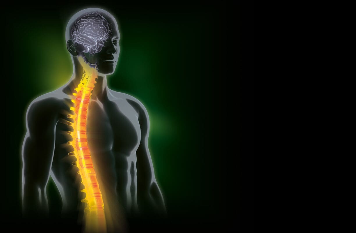
Neuromonitoring in Spine surgery
The spinal cord includes the descending motor nerves that mainly carry impulses from the brain to the muscles, as well as the ascending sensory pathways that transmit information from the peripheral sensory organs to the brain.
There is a general distinction between neuromonitoring of efferent (descending) motor pathways via EMG (electromyography) and motor evoked potentials (MEP), and neuromonitoring of afferent (ascending) pathways using somatosensory evoked potentials (SEP).
What at first glance seems very complex, could be extremely easy with our AVALANCHE® PLUS. In surgical scenarios, where a combination of the different methods is required and measurements are taken simultaneously, AVALANCHE® PLUS has a completely new approach.
Application
Spinal surgery e.g. in scoliosis, kyphosis, decompressions, degenerative spinal diseases, discectomy, during the resection of spinal tumours or for inserting pedicle screws etc.
AVALANCHE® PLUS EMG
for the monitoring of critical proximity to motor pathways
When measuring electromyograms (EMG), motor nerves in the operation site are electrically stimulated. The evoked action potentials are transmitted to the peripheral muscles, then picked up and measured by electrodes and visualized on the neuromonitor.
Continuous EMG recording in several muscles simultaneously enables surgeons to respond immediately when they come critically close to motor neural pathways and signal activity increases.
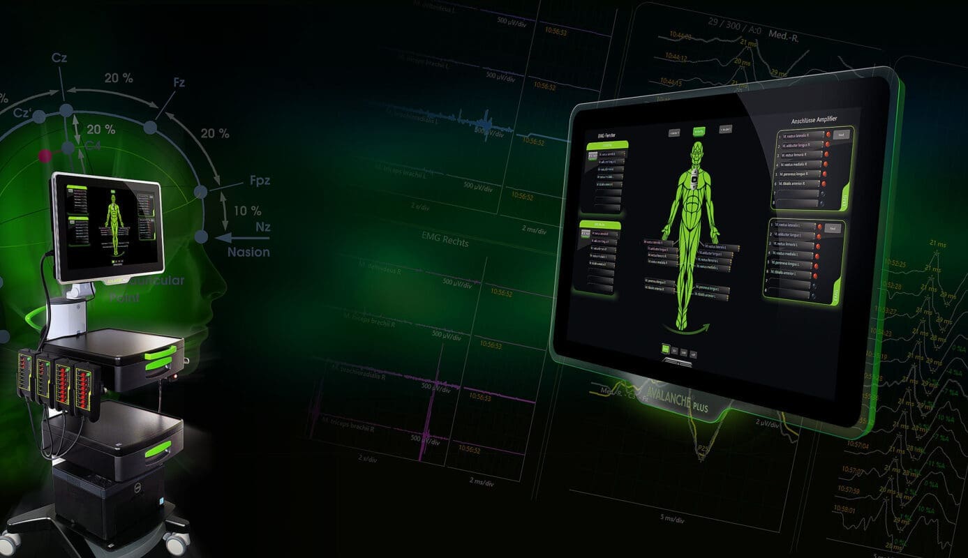
The selection of muscle groups and the required number of channels depends on the surgical scenario. Time consuming entering of muscles via the keyboard is no longer necessary. Setup in AVALANCHE® PLUS is easily done with drag and drop via the touch screen.
AVALANCHE® PLUS comes with predefined muscle groups for every spinal cord level in an anatomic overview. Simply select individual muscle groups and assign them to the desired measuring window. Once you assign the connector, EMG configuration is complete.
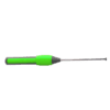
PEDICLE SCREW TESTING
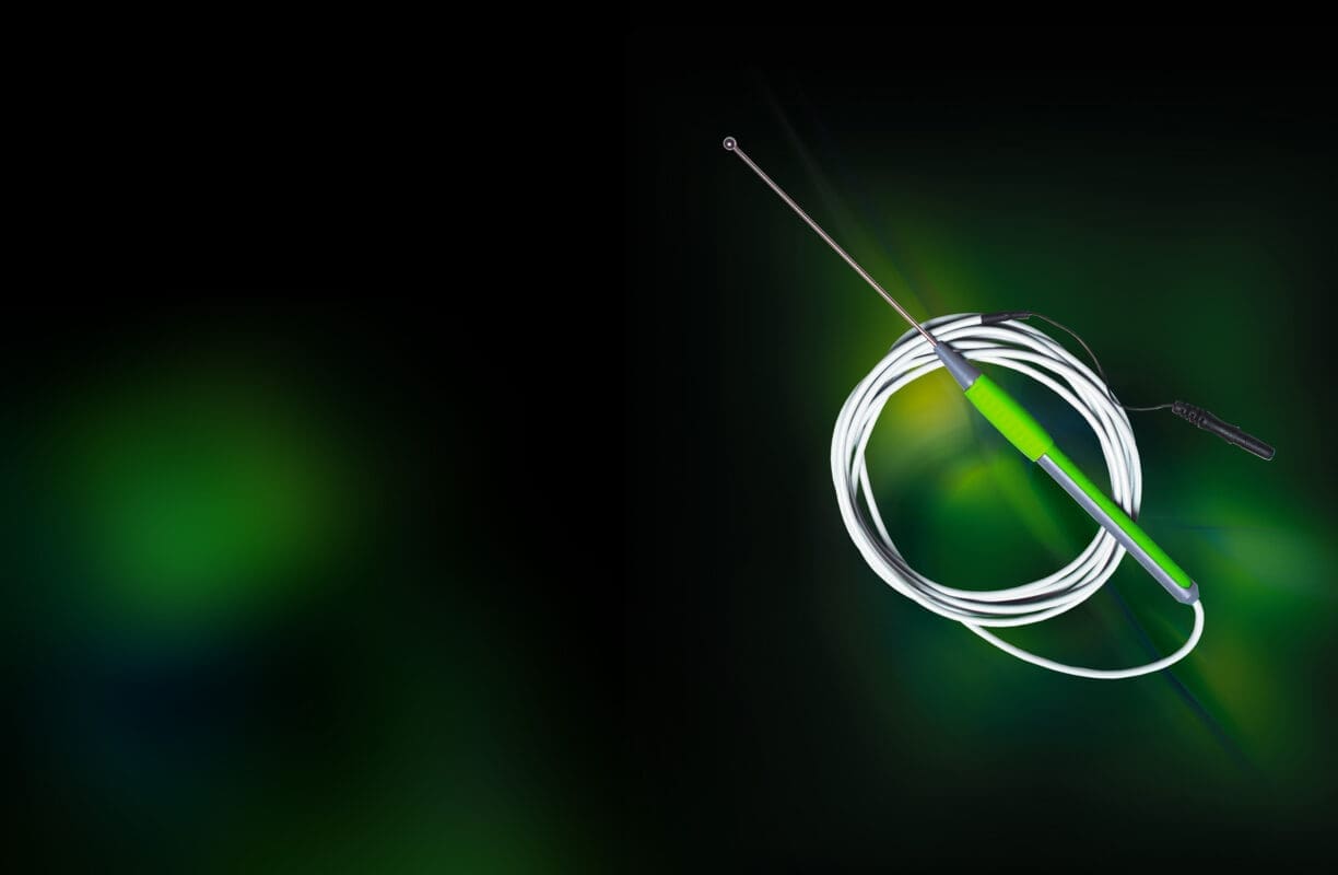
with AVALANCHE® PLUS and the monopolar ball probe
Specific monopolar stimulation probes are available for the insertion of pedicle screws. These permit stimulation within the bore hole itself or through surgical instruments during the drilling and the screwing process. If the stimulation results in an increased signal activity on the neuromonitor, caution is advised – there could be nerve compression or perforation of the pedicle wall.
In the AVALANCHE® PLUS software activation of the pedicle screw stimulator is done by a push of a button. The user interface is identically designed as the AVALANCHE® PLUS I/O-Box. The probe connector on the AVALANCHE® PLUS I/O-Box is colour and shape coded – confusion is nearly impossible.
In the main window you are able to display either all measuring windows of all modalities or one modality only. For monitoring of pedicle screws, choose the EMG view and keep all essential signal changes in view on the large touch screen.
AVALANCHE® PLUS MEP
for the functional check of central motor pathways
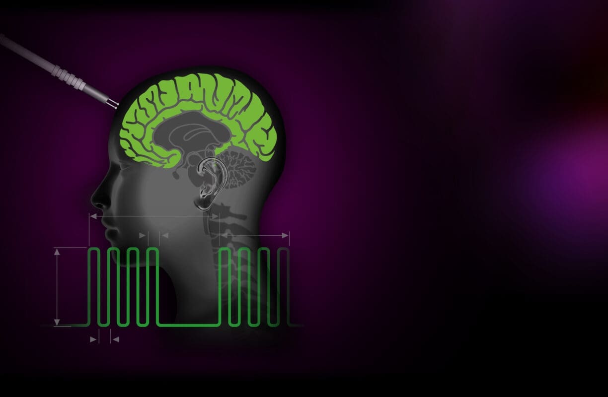
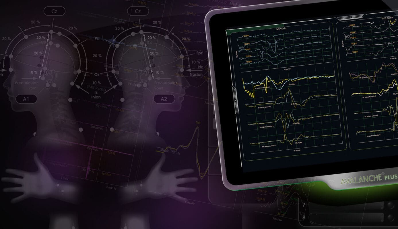
Motor evoked potentials (MEPs) are a particular form of evoked potentials, measured through an EMG. They are triggered by transcranial or direct cortical stimulation, and the response signals are picked up at the corresponding extremity muscles. This enables the surgeon to quickly assess the state and functionality of the spinal cord.
If you have already implemented an AVALANCHE® PLUS EMG setup, MEP configuration is nearly finished. The selection of muscle groups is already done. Just create a MEP measuring window by assigning the muscle groups via drag and drop – setup can be that easy.
If stimulation pulses are released by the transcranial stimulator, AVALANCHE® PLUS triggers the MEP measurement automatically. MEP signals are display in the measuring window without any need of user action. Choose the MEP view to monitor critical phases in surgery comfortable.
AVALANCHE® PLUS SEP
for the monitoring of ascending pathways of the spinal cord
To ensure that changes that could result in post-operative neurological deficits, somatosensory evoked potentials (SEPs) are continually measured. Signals picked up in real time are compared to the normal signal recorded just after anaesthesia was induced – the baseline signal.
The signal generated by electrical stimulation of the median nerve at the wrist or at the tibial nerve at the foot is acquired as an SEP cortically or subcortically and shows typical changes in amplitude and latency. These alert to potential or actual threats for the nerve during operation.
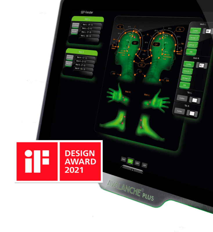
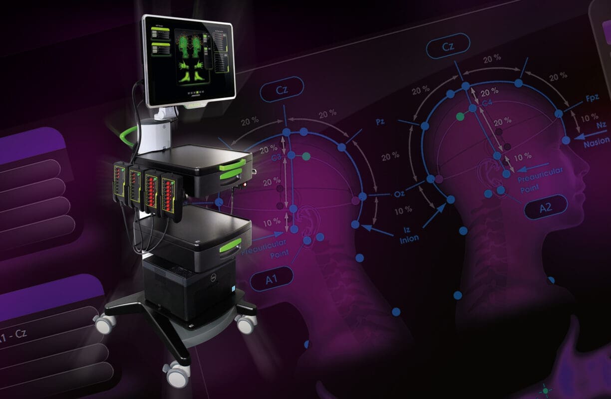
The AVALANCHE® PLUS SEP software comes with predefined SEP channels. At the push of a button, SEP stimulation of the median nerve is chosen. Creating the signal channel manually does no longer exist – AVALANCHE® PLUS creates the signal channel automatically, highlights the required positions of the international 10-20-system in an anatomic overview and selects the stimulation channel by itself.
Assign an electrode connector to the highlighted position of the 10-20 system – easily via drag and drop on the touch screen. In the meantime AVALANCHE® PLUS generated a SEP measuring window to monitor all SEP signals during surgery.
Neuromonitoring in neurosurgery
Intraoperative neuromonitoring of the cranial nerves and central nervous system is routine during neurosurgical interventions.
If a mass makes functionally important areas of the brain unidentifiable during surgery, AVALANCHE® PLUS can help you to find access to the tumour tissue with as little damage as possible, while preserving the brain areas. The function of many cranial nerves can also be controlled with the help of multimodal neuromonitoring
AVALANCHE® PLUS
Multimodal Neuromonitoring
EMG – For testing a critical approach to motor nerves
Monitoring of motor cranial nerves (e.g. cranial nerves III-VII, IX-XII) by means of EMG is essential during surgery on the brain stem and skull base. An approach to motor structures can be detected promptly through continuous recording of muscle activity and acoustic reproduction of the free-running EMG. Stimulated EMG measurements facilitate functional control and localisation of the motor cranial nerves in the surgical site.
SEP – For monitoring sensory pathways and oxygen supply
The continuous recording of somatosensory evoked potentials (SEP) provides important information about the functional state of the sensitive pathways. SEP measurement can, for example, serve to monitor a sufficient oxygen supply to the brain when clipping brain aneurysms – or the posterior funiculus of the spinal cord in the event of intramedullary tumours.
The AVALANCHE® PLUS SEP software offers predefined SEP leads. For example, you can select SEP stimulation on the median nerve at the touch of a button. No manual switching of recording sites required – AVALANCHE® PLUS automatically creates the measuring channel, displays the necessary recording sites in an anatomical view of the international 10-20 system and assigns a stimulation channel. All you need to do is assign the displayed recording site to a connection – quite simply by using the touch screen to drag and drop, while AVALANCHE® PLUS creates an SEP frame in the main window where you can monitor the SEP signals during surgery.
VEP – For monitoring the optic nerve
Intraoperative recording of visual evoked potentials (VEP) is used to monitor the function of the second cranial nerve (optic nerve) and includes monitoring the integrity of the visual pathway from the retina to the visual cortex. Reliable VEP recording can be a support in decision-making regarding the aggressiveness of tumour resection. Real-time adjustment of the strategic approach can reduce permanent, iatrogenic damage to the optic nerve. With AVALANCHE® PLUS VEP, the retina can be stimulated through the closed eye with LED flash goggles (red or white light). The sufficient intensity of the stimulation as a prerequisite for correct VEP measurement can be verified by simultaneously recording an electroretinogram (ERG).
MEP – For monitoring the function of central motor pathways
Measuring motor evoked potentials (MEP) triggered by transcranial or direct cortical stimulation can help monitor and protect motor pathways during almost all neurosurgical procedures by, for example, identifying motor structures or areas of the cortex, or functionally monitoring motor pathways (e.g. pyramidal tracts or motor cranial nerves).
If you have already created an AVALANCHE® PLUS EMG configuration, there is hardly anything left to do for MEP, because the muscles have already been selected. You simply create an MEP measurement frame by assigning the muscles via drag and drop.
AEP – For monitoring the auditory nerve and brain stem
The auditory nerve as part of the vestibulochoclear nerve (cranial nerve VIII) is predominantly located in the cerebellopontine angle. Traumatic stress caused by manipulation around the cerebellopontine angle has a particular impact on this part of the auditory nerve. Maintaining and preserving auditory function is a significant aspect of intraoperative monitoring in cerebellopontine angle surgery, during which the patient is played pre-defined clicking stimuli through earphones.
The resulting action potentials are derived cortically. Automated monitoring of intraoperative changes in AEP potential can help to detect surgical manoeuvres that potentially compromise the patient’s auditory function. AEP potential monitoring provides the surgeon with direct feedback about the functional integrity of the auditory pathway.
DCS – DIRECT CORTICAL STIMULATION
with AVALANCHE® PLUS
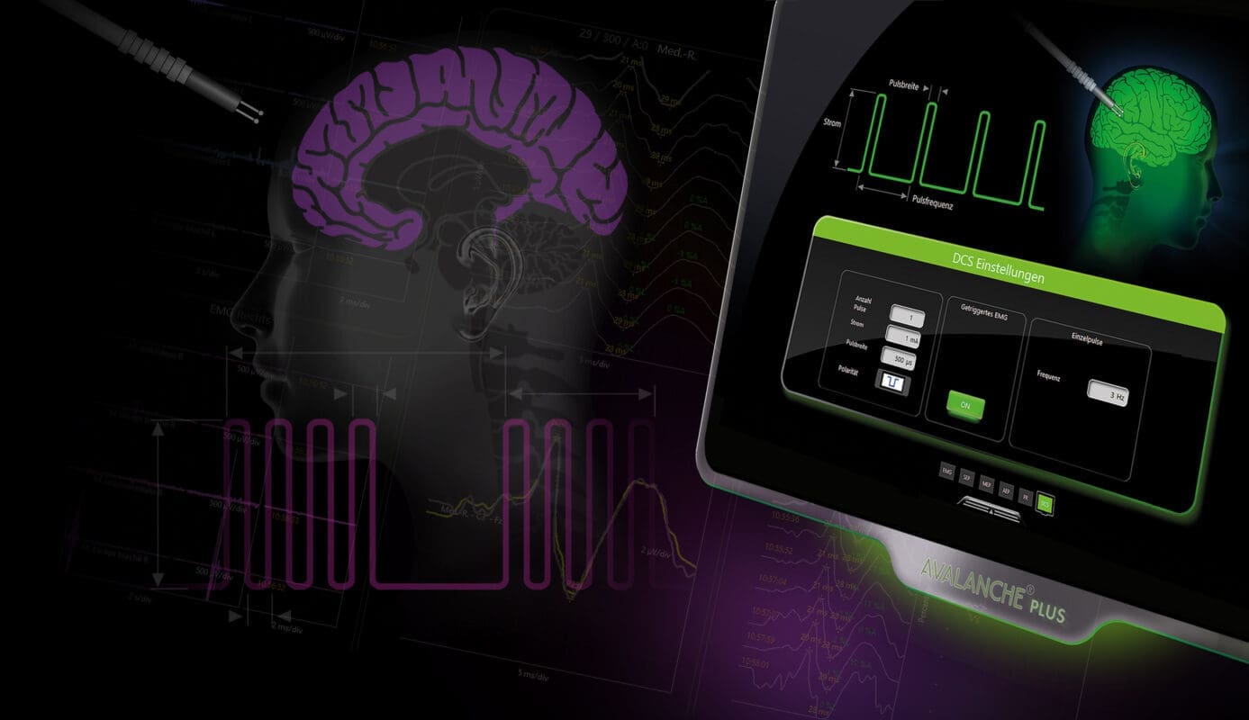
Direct cortical stimulation has been designed for localisation of motor areas in the brain and the speech centre in neurosurgery or epilepsy surgery (cortical mapping). Brain tumours can alter the anatomy of the cortex, which differs from patient to patient in any case, such that, without assistance, access to the tumour tissue with as little damage as possible to the surrounding tissue while preserving functionally vital areas of the brain is difficult. With AVALANCHE® PLUS, it is possible to visualise a “map” of the patient’s individual cortex and localise the regions of interest. Special atraumatic stimulation probes are available for stimulation.
Whether Penfield or train technique, AVALANCHE® PLUS graphically illustrates complex stimulation parameters for direct cortical stimulation. No matter which parameter you change, it is all clarified immediately. Full functionality paired with simplest handling.
AVALANCHE® PLUS makes you a specialist!
SEP PHASE REVERSAL
AVALANCHE® PLUS – Identifying the central sulcus
Tumours in the central cortex can alter the anatomy to such an extent that it is imperative to localise the structures in the area of the central sulcus in order to decide on the type and extent of tumour resection. SEP phase reversal measurements can be used to identify the central cerebral sulcus. The signals usually generated by stimulation at the wrist (median nerve) are measured directly at the cortex with strip electrodes. A polarity change in the evoked potential across the central sulcus can be deduced from the SEP curves, which corresponds to the “phase reversal” between motor and sensory cortex. This can spare healthy tissue and reduce deficits.
Select strip electrodes with 4, 6 or 8 contacts with a single click in SEP phase reversal and let AVALANCHE® PLUS automatically take care of the rest: Electrode visualisation, phase reversal window and phase reversal calculation – complex measurements configured in an instant!

NEURO Surgery
Acustic & Visual Evoked Potentials
-
 Neuromonitor AVALANCHE® SI2 Cirurgia Tiroide
-
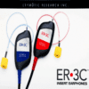
Óculos para VEP
€125.00
-
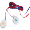
Auriculares para Potenciais Evocados Auditivos
€1,350.00
-
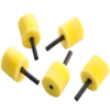
Esponjas AEP para Ouvido
€37.00 – €43.00